- +91 073111 23470
- divakant@gmail.com
- Mon - Sat: 10 AM to 6 PM
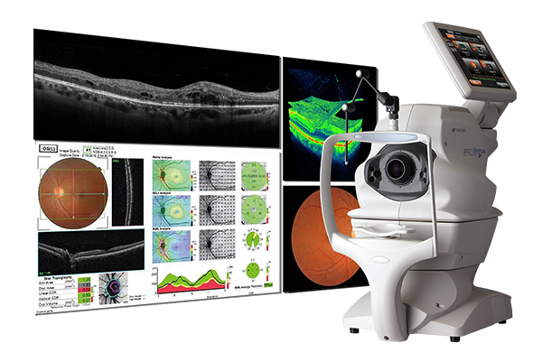
What is optical coherence tomography?
Optical coherence tomography, or OCT, is an imaging method used to generate a picture of the back of your eye, called your retina. The noninvasive method produces an image by measuring the amount of a dim red light that reflects off of your retina and optic nerve. Optical coherence tomography can measure the thickness of your retina and optic nerve.
Healthcare providers of heart and vascular medicine use optical coherence tomography for cardiac catheterization to produce images of your blood vessels. Healthcare providers in the dentistry, gastroenterology, pulmonology, dermatology and oncology fields are also using OCT imaging more often.
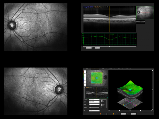
What is optical coherence tomography angiography?
Traditional angiography, also called arteriography, refers to examining the inside of blood vessels using X-rays. Typically, your healthcare provider injects a radiopaque dye that shows up on the X-rays. Eye care professionals also use a special type of angiography to examine blood vessels of your retina. The dye used glows when exposed to a blue light.
Eye care professionals can also use optical coherence tomography angiography to see inside the blood vessels in your eye. Unlike traditional angiography, the test is completely noninvasive and a dye doesn’t need to be injected.
When is optical coherence tomography performed?
Your eye care professional suggests optical coherence tomography if they suspect you have certain conditions at your eye exam or if you already have a condition that they’re helping you manage.
Healthcare providers use OCT to diagnose and manage several conditions that affect the eyes, including:
- Glaucoma: If you have glaucoma, fluid and pressure build up in your eye and damage your optic nerve.
- Age-related macular degeneration: People may lose central vision with this condition. It’s a progressive disease related to aging but, fortunately, treatments are available for some forms.
- Diabetes-related retinopathy: Diabetes damages the small blood vessels of your eye leading to vision loss. Fluid can leak out of your eye causing blurry vision. In severe forms, your entire retina can detach from the back of your eye and glaucoma may develop. People can become completely blind, but with treatment, diabetes-related retinopathy can be controlled.
- Cystoid macular edema: Macular edema refers to the swelling of your macula with fluid. Your macula is the part of your retina that has the most light-sensing cells.
- Macular pucker: Scar tissue can grow over the surface of your retina causing distorted vision. Surgery can sometimes help.
- Macular hole: A macular hole happens when your retina pulls apart forming a hole in your macula. This can affect your vision, but can be repaired by surgery.
- Cone and cone-rod dystrophies: These conditions affect the cells of your eyes that are sensitive to light and color. When the condition worsens, you can have vision loss.
- Tumors in your choroid and retina: These cancers happen in your retina and your choroid, a vascular layer found between your retina and your sclera.
Our Testimonial
I got a retinal detachment surgery done from Dr. Devi Kant Misra at SwarnJyoti Eye Hospital, Aliganj.
The doctor and hospital staff are very responsible and proactive in their efforts.
Highly recommended!
Jaideep Lalchandanipatient
Dr.divakant Mishra sir is very well educated and the most experienced person in the diseases of retina surgery and his nature was very much politeful and been very kind hearted and always be helpful for all of their patients.
Shristi Katiyarpatient
No words for Dr. Diva Kant Mishra. I had a corneal tear in my left eye last month and I'm getting best treatment from Eye Q Hospital, under Diva Kant sir. He listens and watches. He explains everything to patient, so that we can understand deeply. I personally like his check-in notes 😊. He has a very nice and responsible staff also.
Archit Kumar Vermapatient
Previous
Next
